Definition: An opacity in the lens positioned just posterior to the anterior lens capsule and is characterized by the proliferation of anterior lens epithelial cells.
Incidence/Prevalence: Anterior subcapsular cataract is less common than nuclear, cortical, or posterior subcapsular cataract. The subject prevalence for anterior subcapsular opacities is about 1/5 that of posterior subcapsular opacities (Reference 1).
Etiology: There are many associations with anterior subcapsular cataracts including inflammation, trauma, mitotics, amiodarone and atopic dermatitis. In animal models anterior subcapsular cataract has been produced with alkali burns. Some investigators suggest an epithelial to mesenchymal transition in these cells.
Clinical Findings: A focal star-shaped or irregular opacity beneath the anterior capsule as seen with a slit beam.
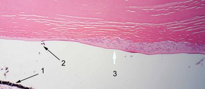 Histopathology: Sections usually show a multilayer of spindled epithelial cells beneath the lens capsule. In the photomicrograph the posterior pigmented layer of the iris is visible (black arrow 1). Previous posterior synechiae to the lens capsule is evidenced by the brown pigment deposition from the iris (black arrow 2). The multilayered lens epithelium is visible (white arrow 3 in figure) and to the left one can see that it is contiguous with a single layer of the normally present anterior lens epithelium. The photomicrograph at higher magnification shows the anterior subcapsular cateract at higher magnifcation, white arrow (the image below has been inverted). Sometimes the anterior lens capsule may be retracted and form slight protrusions.
 Treatment: Surgical removal of the cataract generally restores vision.
References: 1. Ophthalmic Epidemiol. 1997;4;195-206.
