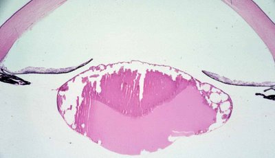
 Histopathology: Histologically, the lens is usually microspherophakic. Frequently, there is accompanying liquefaction and necrosis of lens. The iris is often bowed forward by the lens. In the accompanying gross and microscopic figures, the lens appears small and cataractous. The triangular opacity is prominent anteriorly and seen both in the gross and microscopic examination.
 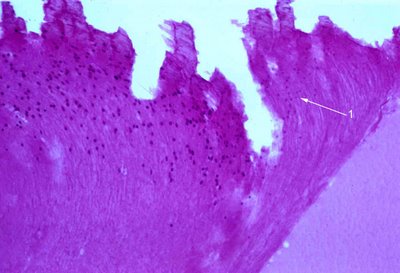 Retained lens fiber nuclei are present. The nuclei may appear pyknotic or karyorrhexic. At white arrow #1 faded nuclei are apparent. Rubella virus may be cultured from surgically removed lenses up to ~3 years of age. PCR techniques may be more sensitive. (Indian J Med Res. 2007 Jan;125(1):73-8)
 A non-granulomatous uveitis affecting particularly the iris has been described by Font and Zimmerman.
Treatment: Prevention through immunization of children and women before they become pregnant is the best approach. Cataract removal can be performed.
