Benign Melanosis of the ConjunctivaDefinition: Pigmentation of the basal layer of the epithelium without a proliferation of melanocytes. This definition includes the ephelis or freckle, so called complexion-associated conjunctival pigmentation, and benign acquired melanosis because the three entities may be indistinguishable clinically and under the microscope.
Etiology: Benign melanosis is a normal phenomenon in heavily pigmented individuals and statistical data suggest that pigmentation may be protective from UV exposure (i.e. dramatically lower incidence of PAM with atypia and conjunctival melanoma). For those with less pigmentation, the etiology is unknown and unknown if such pigmentation is even abnormal.
Clinical Findings: Complexion associated pigmentation occurs mainly at the limbus (arrow 1 in this macrophotograph of an autopsy eye) and fans out to be lighter in color more peripherally toward the fornix
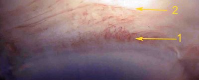
(arrow 2 in the photo) shows that the pigment has dispersed just millimeters from the limbal pigmentation.
Ephelis is a circumscribed lesion.
Histopathology: As shown in the figure, there is increased pigmentation in the cells of the basal layer, i.e. squamous epithelium.
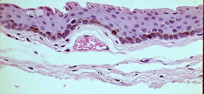
There is no nested pattern and there is no increase in melanocytes. This is critical because the absence of the nesting distinguishes benign melanosis from
primary acquired melanosis. The pigmented granules often are on the apical side of the nucleus as though protection is afforded by absorbing harmful UV radiation. If one looks carefully at the nuclei in the photograph(click on the image to enlarge) one will see the pigment is more concentrated above the nuclei.
Treatment: If recognized in the appropriate setting no treatment is generally needed. However, confusing cases of pigmentation arise that require biopsy. For example if the lesion is actively growing or documented by examination of remote photographs from the patient. In these cases a careful excision should be performed if the lesion is localized and placed flat on filter paper, bottom side down and fixed in formalin. It is very helpful to send a detailed sketch or photo to the eye pathologist to aid in recognizing the clinical problem, orienting the specimen to provide margins and performing careful analysis.
