Fuchs DystrophyDefinition: Fuchs corneal endothelial dystrophy features loss of endothelial cells controlling proper endothelial pump function; thus, malfunction permits marked edema. Vision progressively becomes blurrier and more indistinct as disease advances.
Incidence/Prevalence: Fuchs dystrophy has a variable inheritance pattern and occurs more commonly in women (12.5 to 1.0 in noted familial cases and 4 to 1 in sporadic cases). It is one of the leading causes of bullous keratopathy. Patients rarely present before 30 years and it usually manifests after 40 years of age.
Etiology: Fuchs is genetically inherited from affected parents and has high penetrance as an autosomal dominant disease.
Clinical Presentation: The condition is clinically recognizable by the appearance of guttae in Descemet’s membrane. Focal, anvil-shaped outgrowths are seen buried in the thickened Descemet’s membrane. The outgrowths can also be found protruding into the anterior chamber. The appearance of endothelial cells is sparse or non-existent.
 Histopathology:
 In this disorder the corneal epithelium episodically separates from Bowman's layer and secondary changes occur that are featured here. The epithelial basement membrane reforms from epithelium as it reattaches leading to additional basement membrane material in the middle of the epithelium (number 1). The basal epithelium and sometimes the entire epithelium features a "washed out" appearance (number 2) which on close inspection is seen to be hydropic change. The epithelium is separated from Bowman's layer, a bullous detachment (number 3). Bowman's layer is irregularly thinned (arrowheads 4). The stroma is scarred as seen by irregularly contoured stromal lamellae and an uneven distribution of nuclei within the stroma (difficult to see on this PAS stain). The hallmark of Fuchs dystrophy is of course the guttata that can be seen clinically within Descemet's membrane. In this case the guttata are buried with Descemet's membrane or endophytic (5 and figure below arrows 1). The endothelium is quite sparse but the nuclei are much better seen in a routine H&E stain.
Treatment: Palliative treatment focuses on alleviating symptoms and improving vision after epithelial edema becomes distinct. 5% NaCl drops can be used to clear the epithelial edema or increase tonicity of tear film to prevent accumulation of corneal fluid during sleep. Surgical treatment include transplantion of the cornea or a more limited form of endothelial keratoplasty (DSEK).
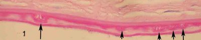
Guttata may be seen clinically with retroillumination at the slit lamp (
see Figure in link).
Usually, guttata in Fuchs dystrophy are exophytic as shown below.
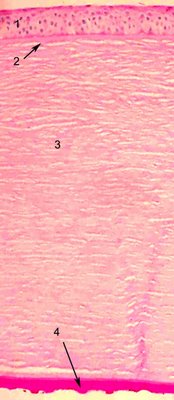
Once again hydropic epithelial changes (1), and irregular Bowman's layer (2) , stromal scarring (3) and expophytic guttata that protrude posteriorly into the anterior chamber (4) are featured. The astute observer will notice that there is pigment in the endothelial cells.
