Choristomas
Definition: Choristomas are defined as mature tissue elements not normally present at the site of occurrence. Examples include limbal dermoid, dermolipoma, ectopic lacrimal gland, and episcleral osseous choristoma. Teratomas require the presence of all 3 germ layers.
Incidence/Prevalence: Choristomas make up about 3% of conjunctival and corneal tumors in some series.
Etiology: Choristomas are congenital proliferations. Choristomas are occasionally familial.
Clinical Findings: Choristomas are the most common epibulbar and orbital tumors in children. Epibulbar choristomas may arise from the cornea, limbus or subconjunctival space, and range in appearance from a small, flat lesion to a large mass filling most of the epibulbar region.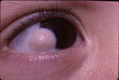 In the photograph the limbal choristoma has extended on to the cornea. Astigmatism is often present. Choristomas may be associated with coloboma, Goldenhar syndrome or epidermal nevus syndromes; those associated with the latter are often bilateral and extensive. Although choristomas most commonly involve the epibulbar area, they can affect many areas of the eye and orbit, and often affect more than one area. Clinically, complex choristomas usually are indistinguishable from dermoids or dermolipomas.
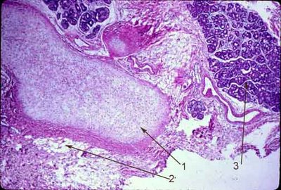 Histopathology: Complex choristomas, in addition to having the features of a dermoid or dermolipoma, include other tissues such as cartilage, bone, and lacrimal gland. In the image above there is cartilage (arrow 1), adipose tissue (arrow 2) and lacrimal gland tissue (arrow 3). A limbal dermoid generally shows a keratinized stratified squamous epithelium covering the surface of the lesion (4) with hair shafts and adnexal structures in the substantia propria. Here sebacaceous glands are prominently displayed (5). In addition the loose collagen of the substantia propria is replaced with dense collagen in thick bundles (6).
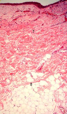
  Compare the limbal dermoid with the dermolipoma (below). The dermolipoma features dense collagen bundles, similar to what is seen in the dermis of skin (7), and adipose tissue (8). However, no adnexal structures are seen. Specifically, neither sebaceous glands nor hair follicles are present in this process. These are the criteria by which this categorical separation is made.
 Treatment: Surgery is generally performed to improve vision or cosmesis, or to impede growth.
