The optic nerve is the bundle of axons that extend from the cell bodies of ganglion cells in the retina to synapse on the lateral geniculate body in the brain. The optic nerve as a structure begins at the optic disk. A smaller, disc-shaped depression, called the cup, lies slightly temporal to the center of the optic disc. On the surface is circular glial plaque, a developmental remnant. Often there is a nasal and inferiorly located Bergmeister’s papilla. Nerve bundles penetrate the collagen of the sclera through a sieve perforations, termed the lamina cribrosa. As the non-myelinated nerves pass this point the optic nerve becomes myelinated; axons are enveloped by a sheath of doubled plasmalemma, to form the myelin produced by oligodendrocytes. Myelination doubles the cross-sectional thickness of the nerve to a diameter of ~ 3 mm at the posterior surface of the sclera. The glial cells of the central supporting tissue meniscus, are astrocytes. The coverings of the optic nerve in the orbit are similar to other brain tissue. The outer most cover
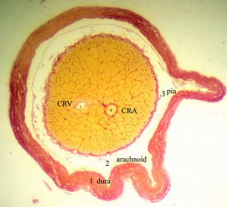
ing of the optic nerve is a sheath of dura. The thick dura mater, fuses distally with the outer layers of the sclera. Internal to the dura is subarachnoid space, arachnoid, and pia. The pia (photomicrograph left) is most internal and tightly applied to the optic nerve proper. Fibrous septa from the pial layer are prominent (shown in red in the photomicrograph above) and enter the optic nerve and divide the axons into fascicles (the yellow bundles in the photo). The central retinal artery (CRA) and vein (CRV) penetrate the optic nerve from 8 to 15 mm behind the optic nerve scleral exit to enter the center of the nerve. The blood supply to the optic nerve is complex, redundant and topographical. Prelaminar or retinal portion is supplied by the short posterior ciliary and recurrent choroidal arteries. The posterior ciliary arteries are terminal branches creating an area that is vulnerable to ischemia. The laminar portion is provided by short posterior ciliary ves
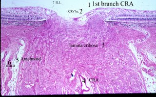
sels via anastomoses with the arterial circle of Zinn-Haller in the sclera. The retrolaminar nerve is supplied by pial, short posterior ciliary, central retinal and perhaps recurrent choroidal vessels. The blood supply for the orbital portion of the optic nerve comes mainly from the ophthalmic artery and a pial network around the nerve. The intracanalicular portion of the optic nerve is perfused entirely by the ophthalmic artery. Drainage of both retinal and choroidal layers appears to be in great part via the central retinal vein and its branches. In the photo ILL refers to the inner limiting lamina of the retina. <
Next Topic in Anatomy>
