What are the boundaries and layers of the iris? The iris, a component of the uveal tract, delineates the anterior and posterior chambers of the eye. The iris contains pigmented cells and muscle and is composed of four layers: the anterior border layer, the stroma, the dilator muscle layer and the posterior epithelium.
The anterior border layer consists of a dense packing of pigmented or nonpigmented cells similar in appearance to the cells present throughout the remainder of the stroma. Absence of cells produces the so called crypts in the border layer.
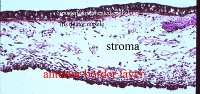
The iris stroma is a loose fibrocollagenous tissue composed of spindle shaped fibroblasts (stromal cells), blood vessels, nerves and macrophages (clump cells of Koganei) containing phagocytosed melanin pigment. At the pupillary margin is the circumferentially arranged smooth muscle of the sphincter muscle of the iris.
What is the blood supply to the iris? The iris is supplied from the major arterial circle that is located in the ciliary body. Rupture of this vessel with ciliary muscle tears is frequently the injury that causes hyphemas. The blood vessels of the iris run in a radial direction. The anterior border layer contains very few vessels. Vessels on the surface of the border layer are abnormal and suggest ischemia in the eye such as in diabetic retinopathy, branch vein occlusions, and neoplasms. Iris blood vessels appeared sheathed and have a characteristic loose appearance. They are nonfenestrated and have pericytes.
What is the composition of the posterior epithelium of the iris? The posterior epithelium is composed of two layers of cells which are densely pigmented with melanin. The posterior boundary of the iris stroma, peripheral to the sphincter muscle, is demarcated by another sheet of smooth muscle, the dilator muscle. The fibers of the dilator muscle are derived from, and remain in continuity with, the cuboidal pigmented cell bodies which make up the anterior layer of iris pigment epithelium.
What determines individual eye color? Eye color is determined by the relative number of melanocytes in the stroma and of course the density of melanin granules produced. Few cells give a blue color, whereas many melanin- containing cells produce a dark brown color; gray and green are the intermediate colors.
It has a circular aperture (pupil) that can be opened and closed by the action of groups of smooth muscle. Contraction of the pupil reduces the amount of light entering the eye and thereby reduces the glare from light scattered from the periphery of the lens.
The dilator muscle layer is composed of the contractile processes of the myoepithelial cells of the inner layer of the posterior epithelium; it extends from the base of the iris to the sphincter muscle.
<
NEXT topic in Ocular Anatomy>
