History: Phacolytic glaucoma was coined in 1955 by Flocks et al. [1].
Etiology: Phacolytic glaucoma is produced by blockage of aqueous outflow by both soluble lens protein and macrophages (as part of the inflammatory response) that obstruct the trabecular meshwork. [2][3]
Incidence/Prevalence: This disorder has markedly decreased in incidence in the U.S. because of earlier cataract extraction. In a large series in India 115 cases of phacolytic glaucoma were reported from 27,073 patients with cataract.[3]. Clinical Findings: Most patients present with pain, a red eye, elevated intraocular pressure, and of course chronic visual loss. The protein and cells will be visualized in the anterior chamber.
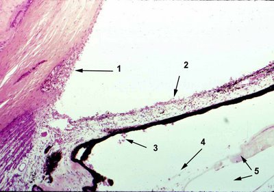
 Histopathology: The proteinaceous material and macrophage accumulate in the angle structures and trabecular meshwork (arrow 1). Macrophages can be found on the anterior (arrow 2) and posterior (arrow 3) surface of the iris, around the lens capsule and lens zonule, (arrow 4), along the inner surface of the retina, and on the optic nerve head. The lens may appear shrunken with loss of nuclear and cortical material and the lens capsule may appear collapsed (arrows 5).
<previous> <next>