Definition- BIGH3 dystrophy is a group of phenotypically distinct corneal dystrophies as distinguished and named by clinical and morphologic characteristics. A single protein appears to be mutated in these dystrophies-keratoepithelin.
Etiology- All of these subtypes arise from mutations in the transforming growth factor beta-induced gene (keratoepithelin) on chromosome 5q31. Over 30 mutations have been described and more are being described all the time. There are 2 hotspots for mutations at Arginine 124 and Arginine 555 of the 683 amino acid 68 kDa protein, known as keratoepithelin. It is not understood how the specific mutations lead to different phenotypes, particularly since the same amino acid can be involved in a point mutation that results in completely different clinical and histologic characteristics. Numerous mutations especially in exons 4, 11-14 have been identified in variants of lattice corneal dystrophy (TGFBI).
Clinical Findings-
Histologic Findings-
Click on the photo to see enlarged view!
Congo red stain shows red-green dichroism with polarized light. The basis of the dichroism is a combination of of optical effects, the strongest of which are dispersion of
birefringence and linear dichroism superimposed on the smaller effects of circular dichroism
and optical rotatory dispersion. There are also fluorescent properties of congo red dye bound to amyloid and the birefringent nature of the protein that rotates the light. Normally white light is transmitted from the microscope thru the slide. A polarizer is placed over the light source that permits only one orientation of light to pass; the light is polarized. Upon illumination with polarized light fluorophores with the absorption transition dipole parallel to the electric vector of the excitation are selectively excited. Congo red is also a fluorophore and absorbs in the green spectral region of wavelengths with a peak absorbance at about ~480-500 nm. The emission (peak 540 nm) is partially polarized as well and there is a Stokes shift. The result is that the fluorescent emission is green (540nm) and polarized. Therefore green will be seen in fluorescence microscopy because it is the fluorescent light but the intensity will be low. However under the light microscope the effect of multiple color in the rotation of the second polarizer called an analyser is due to linear polarized light interacting with dye molecules and the rotation of light (Cotton effect). But red-yellow light will also be seen in some orientation because the amyloid protein has rotated light (the Cotton effect) from the intrinsic birefringence properties and it now has a different orientation. Counterclockwise rotation of the analyzer completely extinguishes the small retardation of the lower wavelengths and long red wavelengths but allows the passage of the wavelengths at the peak retardation. Yellow orange light thus passes the analyzer.
The congo red dye bound to amyloid protein will be seen as either green or yellow or red depending on the orientation of the fibril or the polarizer. Birefringence is characteristic of amyloid, dust, collagen and any crystalline material and so there may be other particles that appear bright. The dystrophies that feature this phenotype are called lattice dystrophy or Avellino dystrophy (the distinction between the two is in fact controversial) but the protein, keratoepithelin, has an expressed point mutation that may be identical to granular dystrophy and Thiel Behnke dystrophy (often misnamed as Reis Buckler's dystrophy) . A litany of mutations have been published but the usual is A

rg124His.
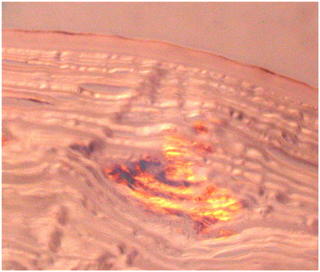
To the left is a photomicrograph of the granular phenotype of BIGH3 dystrophy. Trichrome stain shows the bright red deposit at the arrow that obliterates part of the corneal epithelium and Bowman's layer to lie in the anterior stroma. The proteinaceous deposit is presumably a form of the encoded keratoepithelin protein, but the precise details as to the size, folding and association that permits multiple phenotypes has not been worked out yet.
