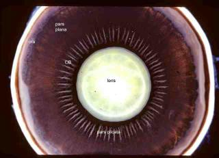
Photograph of a human eye that has been bisected in the coronal plane to show the view of the anterior segment from a posterior perspective (as though you are looking from the retina). The crystalline lens is suspended by delicate fibers called the zonule. The ciliary body (CB) is composed of about 72 processes that make up the pars plicata and a flat area called the pars plana. The ora serrata (ora) is the place where the retina joins the ciliary body. After reviewing the
microscopic sections of an eye with a focus on the anatomy of the cornea your will be ready for images on
corneal pathology.
