Study Guide for dental students and dentists

This is a comprehensive study guide for dental students and dentists. On this site you will Histology of the Human Eye for UCLA Dental Students. In addition there is a comprehensive study guide for the Systemic Pathology.

About Mission for Vision


Sunday, February 26, 2006

Sample Test for Dental students

Here are some sample test questions to help you understand the depth to which you will be examined. Click on what you think is the right answer to confirm. If you think this test is too simple or too difficult let me know by email.

1. Which of the following is least true of the boney walls that surround the eye?
a. the frontal bone forms a portion of the orbital roof
b. the ethmoid bone is thin and prone to sinus infection
c. the lacrimal gland lies in a fossa of the lacrimal bone
d. the floor of the orbit is prone to fracture from trauma
e. the zygomatic bone forms part of the lateral wall of the orbit

2. Which structure is most correctly listed with their functions?
a. lacrimal gland- responsible for secreting lipid to cover the tear film
b. goblet cell- secrete copious amounts of aqueous for the tear film
c. Meibomian gland- secrete mucins to lubricate the eye
d. Gland of Kraus- secrete proteins to prevent infection
e. levator muscle- responsible for closing the eyelds


3. A patient had complications from a recent operation that was performed so he would not need glasses (a LASIK procedure). However, there was a complication and the cornea was made too thin by the laser. During the procedure a laser obliterates tissue from the center of the cornea after a flap is made to peel back the anterior portion (100 microns) of the cornea (normally about 500 microns in central thickness). Given the patient had normal corneal anatomy prior to the operation and knowing the patient's cornea did not rupture, the complication was likely due to overzealous treatment of the:
a. epithelium
b. Bowman's layer
c. epithelial basement membrane
d. stroma
e. Descemet's membrane
f. endothelium


4. Which of the following is least likely to be traversed by a photon traveling to initiate a visual response in the brain?
a. lens nucleus
b. ganglion cell
c. Bowman's layer
d. Bruch's membrane
e. vitreous
f. aqueous


5. A patient has glaucoma because aqueous outflow is slow. A structure adjacent to or connected to the normal pathway has impeded the flow. The LEAST likely cause of the obstruction of aqueous based on anatomic considerations:
a. the peripheral iris became adherent to the cornea.
b. the ciliary body rotated and pushed the lens against the iris.
c. flow across Bruch's membrane was impeded by adjacent abnormal retinal pigment epithelium
d. flow thru the aqueous veins was diminshed by a tumor

6. The cell with the longest axon is the:
a. ganglion cell
b. Muller cell
c. cone
d. rod
e. amacrine cell


7. The vitreous is most firmly attached at the:
a. optic nerve (Bergmeister's papilla)
b. ora serrata (vitreous base)
c. lens (posterior capsule)
d. pars plicata (ciliary processes)


8. The structure in which nuclei are most abundant in the normal adult:
a. anterior lens capsule
b. posterior lens capsule
c. lens nucleus
d. lens equator

9. The structures shown at the tips of the arrows in the photomicrograph function mainly to
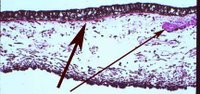
a. modulate fluid (aqueous) transfer
b. regulate an aperature
c. control focus (accommodation)
d. provide structural support


10. Based on the normal anatomy the structure least susceptible to injury from a fist blow to the orbit and eye is the:
a. iris root
b. sclera over the rectus muscle insertions
c. nerves of the inferior orbital fissure
d. intracanalicular portion of the optic nerve

Friday, February 17, 2006

Histology of the Eye for Dental Students

Study Guide for Eye Histology for Dental Students-
Welcome UCLA Dental Students. It is from the material below that questions will be drawn for examination. As you see it is far more complete than is possible in a 50 minute lecture.
The list below is in a question answer format so you can test yourself. Please simply click on the link to go to the answers. Frequently the link forces you to identify the structures. In this way you can further learn histology of the eye. At the end of this list is a sample of examination questions. The choices are linked to information that will enable you to answer the questions.

  1. Name the bones of each orbital wall.

  2. Which orbital bones are prone to blow out fractures and infection?
  3. Name the paraorbital sinuses and their respective locations (and bones).
  4. Which sinus is most relevant to symptoms encountered by a dentist?
  5. What type of epithelium lines the sinuses?
  6. Describe the location and histology of the lacrimal glands.
  7. Name the 3 major proteins that are produced in the lacrimal gland and their role.
  8. What anatomic structures do the tears traverse as they pass from the lacrimal gland cover the eye and exit the orbital area?
  9. Describe the histology of the tear outflow apparatus.
  10. Name the layers of the eyelid and their functions.
  11. Go to the virtual microscope and click on eyelid and study the histology identify the layers of the eyelid including skin, orbicularis muscle, tarsus, meibomian glands, levator, Muller’s muscle and conjunctiva. Your computer becomes a microscope so move the image and change the magnification!
  12. Go to the virtual microscope and click on eye and study the histology.
  13. In a gross photograph of a sectioned eye identify the following structures:
  14. a. conjunctiva, b. cornea, c. anterior chamber, d. lens, e. trabecular meshwork, f. Schlemm’s canal, g. iris, h. ciliary body, i. Pars plana, j. ora serrata, k. retina, l. macula, m. fovea.
  15. Name and describe the layers of the cornea.
  16. Name and describe the layers of the conjunctiva.
  17. What vital component of the tear film is produced by conjunctiva?
  18. Name and describe the layers of the iris.
  19. With contraction which muscle in the iris diminishes and which one enlarges the size of the pupil?
  20. Describe the anatomic structures that make up the anterior chamber.
  21. Follow the flow of aqueous from the cells of production to its exit from the eye.
  22. Name and describe the layers of the lens.
  23. What is structure and composition of the lens zonule?
  24. Describe the gross structure of the ciliary body, pars plana and pars plicata.
  25. Name and describe the layers of the ciliary body.
  26. What is the function of the ciliary muscle in the ciliary body?
  27. What is the structure of vitreous? Where is it most firmly attached?
  28. Name and describe the layers of the retina.
  29. Compare and contrast the histology of the macula, fovea and peripheral retina.
  30. What is the function of the rods? Of the cones?
  31. Describe the process of phototransduction from a histologic point of view (the tissues responsible for each point in the process.
  32. Name and describe the layers of the optic nerve.
  33. What is the artery whose branches supplies all portions of the optic nerve?
  34. Follow a photon of light as it enters the tear film to the final generation of a nerve impulse that synapses in the lateral geniculate body naming all of the structures in between. Sorry no link; it requires integration of all topics.

Sample Questions for the Final Examination!


    Sunday, October 16, 2005

    Anatomy of the Eye- Vitreous

    VITREOUS The vitreous cavity is simply an expanded extracellular space that normally contains 4.0 ml of clear gelatinous substance that is composed largely of water, hyaluronic acid, and collagen. The vitreous normally contains anteroposterior oriented collagen fibrils and occasional macrophages or hyalocytes. The presence of even small numbers of acute or chronic inflammatory cells within the vitreous is distinctly abnormal. The vitreous has distinct attachments to ocular structures. It is most firmly attached attached anteriorly in a circumferential band extending from the posterior pars plana to a few millimeters behind the ora serrata in what has been termed the vitreous base. Traction exerted by the vitreous body at the base results in hyperpigmentation of the underlying pigment epithelium and is evident grossly (Figure 2-1). The vitreous is also attached to the retina over retinal blood vessels and at the optic disc. These attachments are important to understanding vitreous traction, retinal tears, and retinal detachment, for which vitrectomies are sometimes performed.

    Saturday, September 24, 2005

    Pathology of AIDS-HIV-Kaposi

    AIDS-Pathogenesis, Transmission, Diagnosis, PML, AIDS Lymphoma
    1. 1. What is the most common route of transmission of HIV?

    2. 2. What cells in the blood are infected by HIV?

    3. 3. Describe pathogenesis of HIV.

    4. 4. Define AIDS by the CDC criteria.

    5. 5. Describe the geographic distribution of AIDS in the United States and abroad.

    6. 6. State the general categories of scomplications associated with AIDS.

    7. 7. What are the major opportunistic infections seen in AIDS?

    8. 8. Describe the histologic findings of CMV infection.

    9. 9. What are the histologic findings of PML?

    10. 10. Describe the histology of pneumocystis pneumonia.

    11. 11. What is Kaposi sarcoma? Describe its histologic appearance.

    12. 12. What organs are often involved by AIDS related lymphoma?

    13. 13. Describe the neuropathologic features of HIV-1 in the CNS and its manifestations.

    14. 14. What treatment is used for AIDS?

    15. 15. What is the prognosis of patients with AIDS?

    Diabetes Mellitus for Dental Students

    Diabetes Mellitus & Pathology of Diabetes

    • 1.Define diabetes mellitus.

    • 2. What oral pathology lesions are common in Diabetes?

    • 3. What is the incidence of gingivitis and periodontal disease in diabetes?

    • 4. What causes salivary hypofunction in diabetes?

    • 5. Which type of diabetes is more common: Type I or Type II?

    • 6. How many Americans develop diabetes every year?

    • 7. Which ethnic group has the highest prevalence of Diabetes in the United States?

    • 8. What is the relationship between obesity and diabetes?

    • 9. What is the definition of diabetes mellitus?

    • 10. What is the pathogenesis of elevated glucose in diabetes?

    • 11. Describe the pathophysiology of Type I Diabetes and compare it to Type II Diabetes.

    • 12. What are the components of the insulin resistance syndrome?

    • 13. Describe the changes that occur in insulin resistance, with progression of Type II Diabetes.

    • 14. Describe inherited and acquired forms of insulin resistance.

    • 15. List some of the acute symptoms of diabetes mellitus as well as Diabetic ketoacidosis.

    • 16. What are the methods used for diagnosis of diabetes?

    • 17. Describe the similarities and differences between type I and type II diabetes.

    • 18. Give examples of secondary diabetes mellitus.

    • 19. What are some of the ocular complications of diabetes?

    • 20. List some of the ways we can prevent retinopathy.

    • 21. What are the renal complications of diabetes?

    • 22. Describe the renal pathology that can occur in the glomerulus with diabetes.

    • 23. What are the types of diabetic neuropathy?

    • 24. How can we prevent diabetic neuropathy?

    • 25. Describe the general treatment for diabetes.

    • 26. Give examples of drug treatment other than insulin

    • 27. Give examples of insulin analogues of different types.

    Genitourinary Pathology-Kidney-Bladder-Testis-Prostate

    Pathology of the Kidney, Male Genitalia, Cancer of the Prostate

    • 1. Name the three pathogenic mechanisms of glomeruli disorders.

    • 2. Define the clinical and laboratory findings seen in nephrotic syndrome and acute nephritis.

    • 3. Compare the following diseases with respect to age, clinical syndrome of presentation and morphologic changes:

    • 4. minimal change disease

    • 5. focal and segmental glomerulosclerosis

    • 6. membranoproliferative glomerulonephritis

    • 7. membranous glomerulopathy

    • 8. post infection glomerulonephritis

    • 9. IgA nephropathy

    • 10. Goodpasteur’s syndrome

    • 11. What are two important examples of non-immunological glomerular diseases, and what are the clinical manifestations?

    • 12. Name the interstitial renal diseases.

    • 13. What is the most common drug that causes interstitial nephritis?

    • 14. Name the tubular renal diseases.

    • 15. What is the pathogenesis for acute tubular necrosis?

    • 16. Which disease is characterized by inflammation of renal pelvis and interstitium due to acute bacterial infection?

    • 17. Name 3 important malignant neoplasms of the kidney.

    • 18. What is the cellular composition of Wilm’s tumor?

    • 19. What is the histologic appearance of renal cell carcinoma?

    • 20. What is the histologic appearance of transitional cell carcinoma and from what specific sites in the urinary tract are they most likely to arise?

    • 21. What are the two main general categories of lesions in the testes?

    • 22. Which testicular neoplasm is highly malignant, rapidly metastasizing?

    • 23. Which is the most common and least aggressive testicular neoplasm?

    • 24. Cystitis is characterized by what in the urinary bladder?

    • 25. What tumors arise in the prostate?

    • 26. Distinguish the typical clinical presentation of prostate hyperplasia from prostate carcinoma.

    • 27. Describe the histologic findings hyperplasia versus carcinoma of the prostate.

    Female Genital Tract-Gynecologic Pathology

    Uterus, Endometriosis, Endometrial Hyperplasia, Leiomyoma, Venereal Disease etc.

    1. Review the anatomy and histology of uterus.

    2. What are acute and chronic endometritis?

    3. What is endometriosis? Where is it found? What are some of the symptoms? 4. During which period is it most common?

    4. What is endometrial hyperplasia?

    5. What are some of the risk factor that can possibly result in endometrial hyperplasia?

    6. What are some histological findings of endometrial hyperplasia?

    7. What is endometrial carcinoma?

    8. List the staging categories of endometrial carcinoma and the relationship to prognosis.

    9. Compare the presenting findings and morphology of leiomyoma and leiomyosarcoma.

    10. Define pelvic inflammatory disease. What are its symptoms?

    11. How is gonorrhea transmitted? What are the symptoms of infection? What are some of the complications of this disease?

    12. What are some of the conditions attributed to chlamydia trachomatis in both women and men?

    13. Where is the site of infection of herpes simplex virus type I & II? What are the complications?

    14. Describe the stages of syphilis infection.

    15. What does recent molecular and epidemiological data suggest about the etiologies of mild dysplasia/CIN I? moderate and severe CIS, CINII and CIN III?

    16. List some of the significant risk factors for cervical dysplasia and neoplasia.

    17. Name the common neoplasms in the following sites: vulvar,vagina, cervix, uterus.

    18. What treatments are available for squamous dysplasia and carcinoma of the cervix?

    19. Describe the stages of cervical cancer and the approximate survival rate at each stage.

    20. What are five types of ovarian tumors?

    21. Describe the stages of ovarian carcinoma and the approximate survival rate at each stage.

    Hematopathology Lymphoma-Blood and Bone Marrow Pathology

    Hematopathology

    1. 1. Define anemia. What are some of the general ways anemia may occur?

    2. 2. Define myelodysplastic syndrome.  What is the major complication of this syndrome?

    3. 3. Name the 4 major classes of myeloproliferative disorder.

    4. 4. Define leukemia.

    5. 5. Define malignant lymphoma. Name the 2 major categories and how they are distinguished in terms of the types of cells that proliferate.

    6. 6. What are monoclonal gammopathies? Give two examples.

    7. 7. Define hemostasis.

    8. 8. Name some clinical manifestations of thrombocytopenia.

    9. 9. Name the major diseases that involve coagulation factor defects.
    • 10. Name 3 disorders that promote blood clotting and describe their pathogenesis.

    Pathology of Bone Including Tumors for Dentists

    Pathology of Bone

    • 1. List the types of bone found in our body.

    • 2. Describe the morphologic features of woven bone.

    • 3. Describe the morphologic features of lamellar bone.

    • 4. What information is important to be able to diagnose bone tumors?

    • 5. Give the age group, common location, morphologic characteristics, and general treatment of:

    • 6. Fibrous dysplasia

    • 7. osteochondroma

    • 8. enchondroma

    • 9. osteoid osteoma

    • 10. osteoblastoma

    • 11. benign giant cell tumor of bone

    • 12. primary chondroblastoma

    • 13. chondrosarcoma

    • 14. Ewing’s sarcoma

    • 15. chordoma

    • 16. Name the most common bone tumors that metastasize to bone.

    • 17. Define the x-ray findings, distribution, and histologic appearance of multiple myeloma.

    Diseases of the Hepatobiliary Tree-Pancreas

    Liver, Biliary Tree, Pancreas

    1. 1. Review the anatomy and histology of the liver, pancreas, and gallbladder.

    2. 2. Review the circulation of the liver.

    3. 3. Describe the patterns of necrosis found in pathology of the liver.

    4. 4. What is the significance of ballooning degeneration of hepatocytes?

    5. 5. What are the morphologic features that define cirrhosis of the liver?

    6. 6. Differentiate the etiology, route of spread and morphologic characteristics of Hepatitis A, B and C.

    7. 7. What are the current treatments for Hepatitis B?

    8. 8. Name the risk factors for hepatocellular carcinoma.

    9. 9. How does one distinguish hepatocellular carcinoma from normal liver?

    10. 10. What disease accounts for the vast majority of biliary tract disease worldwide?

    11. 11. What is the pathogenesis of acute pancreatitis?

    12. 12. What are some of the risk factors for pancreatic adenocarcinoma?
    • 13. Describe the clinical features of pancreatic adenocarcinoma.

    Diseases of the Breast

    Breast Enlargement, Fat Necrosis, Fibrocystic Disease, Breast Cancer

    1. 1. Describe the anatomic features of the female breast.

    2. 2. What causes the enlargement of breast during pregnancy?

    3. 3. List some of the congenital diseases of the breast.

    4. 4. Describe the histologic features of acute mastitis.

    5. 5. Describe the histologic features of fat necrosis.

    6. 6. What is the most common disease of the female breast?

    7. 7. What breast lesions are amenable to diagnosis by fine needle aspiration?

    8. 8. What are the pathologic findings seen in fibrocystic disease of the breast?

    9. 9. What is the relative risk of cancer in fibrocystic disease of the breast?

    10. 10. What is the clinical presentation of fibrocystic disease of the breast?

    11. 11. What is the typical clinical presentation of a fibroadenoma?

    12. 12. What are the key histologic elements of a fibroadenoma?

    13. 13. What are the risk factors for breast cancer?

    14. 14. What are the receptors of particular prognostic importance in breast cancer?

    15. 15. Describe the role of the BRAC 1 and 2 gene mutations in breast cancer.

    16. 16. What are the advantages of mammography?

    17. 17. Describe palpable mass and non-palpable mass.

    18. 18. What is the distribution pattern of cancers of the breast compared to benign lesions?

    19. 19. What are the routes of spread of breast cancer?

    20. 20. Describe the pathologic features of breast cancer. What is the most common type?

    21. 21. What operations are currently performed for breast cancer?

    22. 22. What chemotherapeutic agents are currently used and under what circumstances?

    23. 23. What is the role of radiation for breast cancer? (When is it used?)

    24. 24. What factors influence survival for breast cancer?

    25. 25. What is the definition gynecomastia of the male breast? What treatment is undertaken?

    Central Nervous System and Muscle

    Arnold-Chiari malformation, hydrocephalus, meningitis, neoplasms

    1. 1. What abnormalities are associated with errors of fusion of the bony spinal canal?

    2. 2. Describe Arnold-Chiari malformation.

    3. 3. What is Hydrocephalus?

    4. 4. List the three forms of CNS hemorrhage associated with trauma? Describe each form and the pathogenesis.

    5. 5. What two complications occur as a result of compound fractures to the skull?

    6. 6. What are the four predisposing circumstances leading to infections of pyogenic, viral or granulomatous disease?

    7. 7. What causes meningitis in the elderly?

    8. 8. List at least three examples of granulomatous infection.

    9. 9. Define progressive multifocal leukoencephalopathy.

    10. 10. How do tumors produce signs and symptoms? Describe two basic mechanisms.

    11. 11. Define* glioma (astrocytoma).

    12. 12. Define* oligodendroglioma.

    13. 13. Define* medulloblastoma.

    14. 14. Define* meningiomas.

    15. 15. Define* Schwannoma.

    16. 16. Define Congenital aneurysms.

    17. 17. What are some of the causes of intracranial hemorrhage?

    18. 18. Describe cerebral infarcts.

    19. 19. What is meant by demylinating diseases? Give an example.

    20. 20. What causes Wernicke's encephalopathy?

    21. 21. Describe the clinical and pathologic features of Tay-Sachs disease.

    22. 22. What are two major microscopic changes seen in Alzheimer's disease?

    23. 23. What are various categories of myopathies?

    Respiratory Disease

    Chronic Obstructive Pulmonary Disease, Bronchiole Types, Pulmonary lobule, Collateral Ventilation, Emphysema, and Lung Disease

    • 1. What is COPD, and what is it characterized by?

    • 2. Provide a description of bronchi, and differentiate them from the bronchioles.

    • 3. What are the types of bronchioles?

    • 4. What is the acinus?

    • 5. What is the pulmonary lobule?

    • 6. Describe collateral ventilation.

    • 7. What defines emphysema? Describe its pathology.

    • 8. Compare centrilobular and panlobular emphysema.

    • 9. List and describe the major pathologic changes that occur in COPD.

    • 10. What are some clinical syndromes of COPD? How are they clinically defined?

    • 12. What are some causes of airflow limitation?

    • 13. What is the proteolytic enzyme hypothesis?

    • 14. What are some ways in which lung cancer is related to smoking?

    • 15. Define squamous cell carcinoma, adenocarcinoma, small cell undifferentiated carcinoma, large cell undifferentiated carcinoma, bronchial adenoma, and mesothelioma.

    • 16. Which of the types of lung cancer are least likely to present as central masses?

    • 17. List the five ‘paraneoplastic’ syndromes.

    • 18. What are the tests used to diagnose a lung tumor?

    • 19. What are possible treatments, and when should each be used?

    • 20. What are the determinants of prognosis in lung cancer?

    • 21. Which type of lung cancer has the best overall prognosis? Worst?

    Cardiac and Vascular Diseases

    Cardiac and Vascular Diseases

    • 1. What are the various classification schemes for Heart Disease?

    • 2. Describe the pathophysiologic classification.

    • 3. Describe the structural classification

    • 4. Describe the classification by pathogenesis.

    • 5. Compare congenital versus acquired heart disease, and give examples of each.

    • 6. Name the categories of complications from cardiac valvular defects.

    • 7. Compare and contrast the causes and effects of left-sided heart failure and right-sided heart failure.

    • 8. Define shock. Provide some examples. Which is most commonly seen by dentists?

    • 9. How are the three stages of shock characterized?

    • 10. How is neurogenic shock treated?

    • 11. Name the symptoms that may develop form anaphylaxis. How can it be treated?

    • 12. Define hypertension.

    • 13. What is the incidence of hypertension?

    • 14. What symptoms does hypertension produce in most people with the disease?

    • 15. What proportion of African American adults has hypertension?

    • 16. What are the four categories of diseases associated with hypertension?

    • 17. How can hypertension be controlled?

    • 18. Define hyaline arteriolosclerosis.

    • 19. Name two types of malignant hypertension.

    • 20. What are the complications of hypertension?

    • 21. Define atresia, and stenosis.

    • 22. What is the pathogenesis of endocarditis?

    • 23. What dental and surgical procedures predispose to bacteremia?

    • 24. How is primary myocardial disease characterized?

    • 25. How does pericardial disease cause heart disease?

    Thrombosis and Embolism

    Platelets, Prostaglandins, Coagulation, Thrombosis, Hematoma
    • 1. What are the elements involved in hemostasis, and what is most important?

    • 2. Describe the function of platelets.

    • 3. What are prostaglandins and what do they do?

    • 4. In what tissues are prostaglandins commonly found?

    • 5. How are prostaglandins made by the cell?

    • 6. Describe what can affect the coagulation system.

    • 7. Define hematoma.

    • 8. What is thrombosis? Describe its pathology.

    • 9. Compare white and red thrombi.

    • 10. What is embolism?

    Morphology and Behavior of Tumors

    Dysplasia, Neoplasms, Cancer, Staging, Differentiation

    • 1. Define differentiation. What is the difference between well-differentiated and poorly-differentiated cancer cells?

    • 2. Differentiate dysplasia from cancer.

    • 3. Define tumor.

    • 4. What is a neoplasm?

    • 5. Compare the rate of growth in benign and malignant neoplasms.

    • 6. Compare invasion in benign and malignant tumors.

    • 7. What is metastasis and how does it define a tumor?

    • 8. What are the routes of metastatic spread?

    • 9. What is the difference between tumor grade and tumor stage?

    • 10. Describe dysplasia.

    • 11. Compare carcinoma versus sarcoma.

    • 12. State the correct terminology for: a) benign glandular tumor b) malignant

    • glandular tumor c) benign mesenchymal tumor of fat d) malignant mesenchymal

    • tumor of fat

    • 13. Define the features that are indicative of malignancy.

    • 14. Define the features that are indicative of benignity (neoplasm).

    • 15. Define the terms, pleomorphism, and anaplasia.

    • 16. What is the significance of abnormal mitotic figures being present in neoplasm?

    • 17. Compare a stage I malignant neoplasm with a stage IV malignant neoplasm.

    • 18. What are features that relate to architectural abnormalities in histologic section ?

    • 19. What are features that relate to cytologic abnormalities in histologic section ?

    Biology and Biochemistry of Cancer

    Cancer Incidence, Mortality Rate, Effect of Age, Etiology (Cause)
    • 1. List and describe the events of cancer progression.

    • 2. The properties of cancer can be studied by what methods?

    • 3. Compare and contrast properties of neoplastic cells versus normal cells.

    • 4. Define cancer incidence and cancer mortality.

    • 5. What is the relationship between age and cancer?

    • 6. What cancers are the most prominent causes of death in men? In women?

    • 7. Is the incidence of death from cancer rising or decreasing?

    • 8. What is the most likely general cause of cancers, worldwide?

    • 9. Define protooncogenes and tumor suppressor genes. What are their normal functions?

    • 10. Name the types of mutations involved in altering these genes.

    • 11. Name two tumor suppressor genes.

    • 12. Describe how cancer can occur by genetic change

    • 13. List the three classes of agents to cause cancer. For each class, describe the ways in which cancer can be caused.

    • 14. What are some factors that can increase the incidence of tumors?

    • 15. Name a specific type of cancer and the risk factors for that type.

    • 16. What are some ways in which cancer can be managed?
    17. What are the risk factors for lung cancer? Breast cancer? Uterine cancer? Colon cancer? Oral cancer?

    Wound Healing

    Non-specific wound healing (general concepts); Specific Wounds, Healing by Primary and Secondary Intention.

    • 1. Describe the sequence of events in non-specific wound healing, and provide a related time table.

    • 2. Define wound contraction.

    • 3. Define wound strengthening.

    • 4. Compare and contrast abrasion from incision/laceration.

    • 5. Describe a wound with epidermal defects.

    • 6. Which substances are elaborated, and by which cells (i.e., collagen types, fibronectin, etc)?

    • 7. Compare temporary matrix composition vs. definitive matrix composition.

    • 8. List the cells, and cell components involved in wound contraction. Provide a time frame of events.

    • 9. What is the tensile strength of the wound at time points 1 week, and 3 months?

    • 10. Compare the tensile strength of various collagens.

    • 11. Describe the time course of healing of a sutured skin wound.

    • 12. Compare the processes of primary vs. secondary intention in terms of inflammation, necrosis, cellular proliferation, and scar formation.

    • 13. What are the events of healing following bone fracture?

    • 14. What are the growth factors involved in wound healing?

    • 15. List the local and systemic factors that interfere with wound healing.

    • 16. Define pyogenic granuloma and keloid, and describe their morphologic features in histologic sections.

    • 17. What is the role of matrix metalloprocesses in wound healing?

    • 18. Name a few diseases and factors that interfere with healing.

    Biology and Biochemistry of Cancer page 6

    Cancer-Incidence-Cancer Genetics-Protooncogenes-Suppressor Genes

    • 1. List and describe the events of cancer progression.

    • 2. The properties of cancer can be studied by what methods?

    • 3. Compare and contrast properties of neoplastic cells versus normal cells.

    • 4. Define cancer incidence and cancer mortality.

    • 5. What is the relationship between age and cancer?

    • 6. What cancers are the most prominent causes of death in men? In women?

    • 7. Is the incidence of death from cancer rising or decreasing?

    • 8. What is the most likely general cause of cancers, worldwide?

    • 9. Define protooncogenes and tumor suppressor genes. What are their normal functions?

    • 10. Name the types of mutations involved in altering these genes.

    • 11. Name two tumor suppressor genes.

    • 12. Describe how cancer can occur by genetic change

    • 13. List the three classes of agents to cause cancer. For each class, describe the ways in which cancer can be caused.

    • 14. What are some factors that can increase the incidence of tumors?

    • 15. Name a specific type of cancer and the risk factors for that type.

    • 16. What are some ways in which cancer can be managed?

    • 17. What are the risk factors for lung cancer? Breast cancer? Uterine cancer? Colon cancer? Oral cancer?

    Hereditary and Congenital Disease page 5

    Inborn Errors of Metabolism, Down's Syndrome, Cri Du Chat, etc.

    • 1. What are the three categories of hereditary diseases? Describe each.

    • 2. Why are Mendelian disorders often called inborn errors of metabolism?

    • 3. What are some ways in which chromosomal diseases can occur?

    • 4. Recognize the pattern of transmission from a detailed family tree (i.e., autosomal dominant, autosomal recessive, X-linked etc.).

    • 5. Predict the likelihood of recurrence in another offspring in diseases with the transmission of inheritance as evident in the family tree.

    • 6. Describe the pathogenesis of phenylketonuria, and its mode of transmission.

    • 7. Describe how inborn errors of metabolism affect substrate fate

    • 8. Why are autosomal trisomies usually always incompatible with life?

    • 9. Describe how the individualiazation (visualization) of the chromosome was achieved.

    • 10. What are some diseases diagnosed by cytogenetics?

    • 11. Define translocation and explain its role in Down’s syndrome and Cri du Chat.

    • 12. What is karyotyping?

    • 13. Provide a description of the effects of Klinefelter’s Syndrome.

    • 14. What is the Lyon hypothesis?

    • 15. Describe the ‘ethical issues’ of genetic testing.

    • 16. Define mosaicism.

    Infectious Processes page 3

    Host Defense- Bacteriocins, Bacterial, Fungal, Viral Infections

    1. Name the determinants of pathogenicity.

    2. Recognize host defense mechanisms.

    3. Give an example of mechanical barriers.

    4. Define bacteriocins.

    5. Name some organisms that can penetrate the skin.

    6. Name some organisms that can invade mucous membranes.

    7. How do tears and mucous protect against infection?

    8. What is the effect of exogenous steroid hormone on the risk of infection?

    9. Explain how Gram negative bacteria can cause fever.

    10. How do endogenous pyrogens affect temperature regulation?

    11. What are some non-specific and specific factors that increase susceptibility of the host to infection?

    12. What is the A-B model?

    13. What is the human lethal dose of botulinum toxin type A?

    14. Name the most common pathogenic fungi.

    15. What percent of fungal types are pathogenic in humans?

    16. Approximately how many pathogenic viruses exist?

    17. What is the proportion of bacteria that are human pathogens?

    18. List the types of parasites that can cause disease.
    What is the worldwide percentage of parasitic disease infection?

    Inflammation

    Inflammation - Cells, Mediators & Immune and Autoimmune Disease

    • 1. List the changes that occur in hemodynamics, vascular permeability and inflammatory cell movement in acute inflammation

    • 2. What are the three types of granulocytes?

    • 3. Describe the components of the reticuloendothelial system.

    • 4. Define acute inflammation, chronic inflammation, and sub-acute inflammation.

    • 5. Define the term granuloma. Give an example of a disease characterized by granulomata.

    • 6. List and briefly describe some of the cardinal signs of inflammation.

    • 7. What is the complement system? Describe the two pathways.

    • 8. What is histamine?

    • 9. What can cause histamine to be released into the surrounding tissues?

    • 10. What are the effects of histamines?

    • 11. What is bradykinin? Describe how it is activated.

    • 12. What are prostaglandins?

    • 13. What are lymphokines?

    • 14. Define neutrophilic enzymes and their role in inflammation.

    • 15. Describe the similarities and differences among the four types of hypersensitive reactions.

    • 16. Describe the immune pathogenesis of each of the following:

    • 17. Goodpasture’s disease

    • 18. erythroblastosis fetalis

    • 19. pernicious anemia

    • 20. lupus erythematosus

    • 21. rheumatoid arthritis

    • 22. scleroderma

    • 23. Name four types of hypersensitivity reactions, and give a practical example of each.

    Study Guide for Dental Students- Cell Growth and Regeneration

    Embryoblast, trophoblast, cleavage, hypertrophy, atrophy, hyperplasia etc.

    • 1. What is an embryoblast?

    • 2. What is a trophoblast?

    • 3. What is a zygote?

    • 4. Define cleavage.

    • 5. What are the 3 germs cell layers, and what types of tissues does each of the layers become?

    • 6. What structures have a phospholipid bilayer?

    • 7. What is the composition of the ribosome?

    • 8. What is the cytoskeleton and where is it located?

    • 9. What are some types of fibers found outside of the cell?

    • 10. What are two common stains used to dye RNA, protein, and elastic fibers?

    • 11. List the four types of tissues and where each type is found.

    • 12. List the three categories of cells based on their division. Describe each.

    • 13. Define hypertrophy.

    • 14. Define atrophy.

    • 15. Define hyperplasia.

    • 16. Define dysplasia.

    • 17. Define metaplasia.

    • 18. Define hydropic swelling.

    • 19. Compare and contrast the features of reversible vs. irreversible cell injury.

    • 20. Define pyknosis, karyorrhexis, and karyolysis.

    • 21. What is apoptosis? List the events that occur from apoptosis.

    • 22. Define the three types of necrosis: coagulative, liquefactive, and caseous.

    • 23. Give one example for each type of necrosis.

    Dental Student Guide for Systemic Patholoogy


    • TABLE OF CONTENTS-STUDY GUIDE FOR DENTAL STUDENTS AND DENTISTS-SYSTEMIC PATHOLOGY